Diagnosis
What is the diagnosis?
The diagnosis is ALK-positive large-cell T-cell lymphoma.
T-Cell lymfoma?
T-Cell lymphoma I have cancer of the T-cells in my lymph nodes (in this case, in my right armpit). These cells are part of my immune system and live in my lymph node. They have started rapidly dividing in there after a random mutation and have formed a tumour. This is a malignant tumour, meaning that it will spread to other parts of my body if given the chance.
Is that hodgkin-lymphom?
No, this is a much rarer non-hodgkin lymphoma.
ALK-positive?
ALK-positive means that the outside of the cancerous cells has a specific protein. This is a protein modern chemotherapy can bind to. This means the chemotherapy can work more effectively and more targeted.
How were you diagnosed?
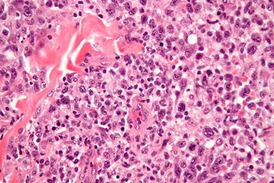
The diagnosis was made by the anatomical pathologist in the Reinier de Graaf Hospital in Delft in Holland, and verified by the pathologist in the Leiden University Medical Center. They have come to the diagnosis by testing a small sample of my lymph node removed though a cut in my armpit. The pathologist adds a dye to the sample to be able to see the cells well. They then look at the sample under a microscope to determine what types of cells they are. Such a stain looks like this:

How far advanced/in what stage are you?
There are four stages to lymphoma:
- There is lymphoma in one group of lymph nodes or in one organ.
- There is lymphoma in 2 or more groups of lymph nodes or in one organ.
- There is lymphoma in lymph nodes above and below the diaphragm.
- The lymphoma has spread beyond the lymph nodes into the liver, spleen, bone-marrow, or the lungs.
I had a PET- and CT-scan the 4th of January 2013 to investigate where the lymphoma was. Despite a somewhat unclear result the oncologist carefully categorised me in phase-1. My bone-marrow and bone were investigated and turned out to be clean. This was tested by inserting a large needle into my pelvis (breaking the bone) and sucking out some bone-marrow. This was the least pleasant test of all.
Unclear?
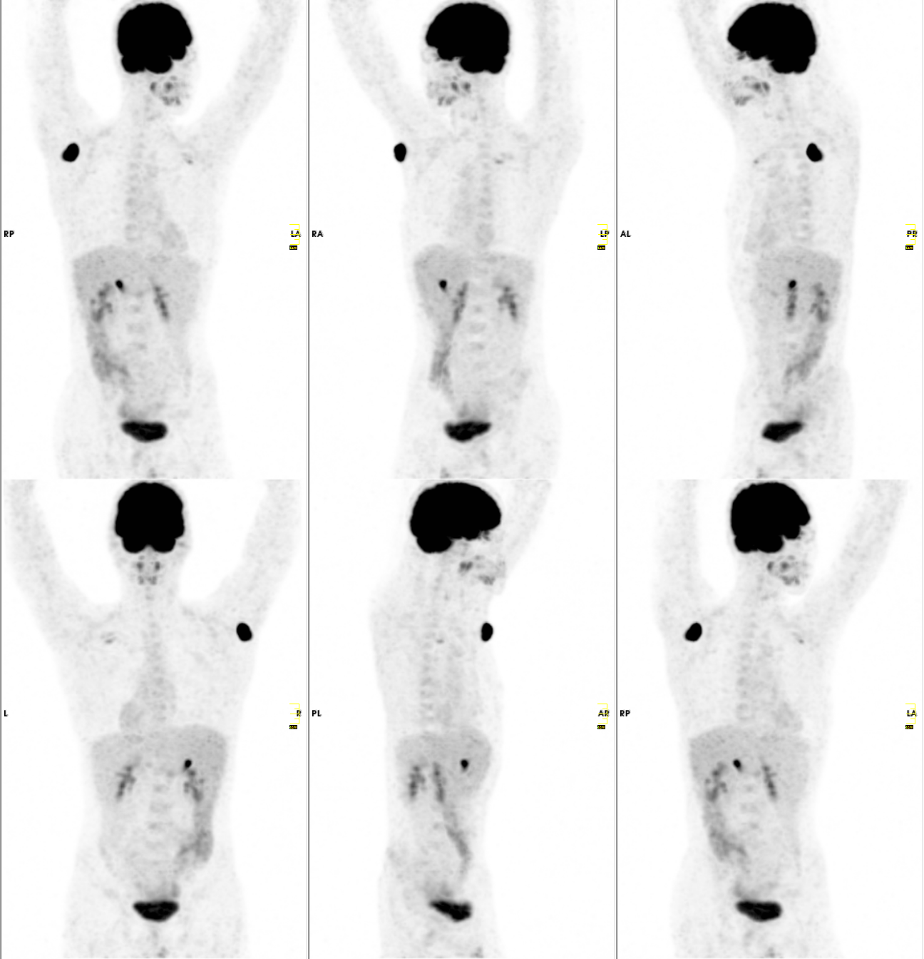
Yes, the magic word is uncertainty. The PET scan wasn’t completely clear. My gallbladder did light up, and a spot was visible behind my left pectoral muscle. When I went to get the PET scan I wasn’t allowed to eat for 6 hours. I was then injected with glucose, with a radioactive marker bound to it. Because my body had not had any food for a long time and was keen for energy it started to use the glucose with the radioactive marker attached to it. The spots in my body that used the most glucose (like a hungry tumour) absorbed the largest amount of the radioactive marker. The scan looks at the spots in the body that emits the most radiation and uses this to construct a 3D image. On the scan you can see that my brain is lit up because it uses a large amount of energy. My kidneys and bladder light up because they filter some of the substance out of my blood. My gallbladder lighting up could be because of the cancer, but it would be quite uncommon. The radiologist I spoke to about this suggested it could possibly be an infection, or my gallbladder happened to be active during the time the marker was injected. A second scan halfway through my treatment (in March) should give more clarity.
The scan
This is an animation of my PET scan. Because it looks kind of cool and gives you an idea of what I’m on about.
